Visceral Adipose Tissue: How Organ-Surrounding Fat Differs Metabolically From Subcutaneous Fat
Visceral fat surrounds the organs and is metabolically distinct from subcutaneous fat under the skin. Learn why location matters, how CT and MRI measure it, and what waist circumference cannot tell you.
Your Waist Is Hiding a Metabolic Time Bomb
The fat you can pinch under your skin is not the fat that damages your organs. Two people with the same total body fat can have completely different health outcomes. Visceral adipose tissue surrounds the liver, pancreas and intestines inside the abdominal cavity. Subcutaneous fat sits beneath the skin outside the muscle wall. Location determines the danger. Visceral fat drains directly into the portal vein, releasing free fatty acids and pro-inflammatory adipokines straight to the liver. That is why a person with a flat stomach can have dangerous visceral fat and a person with pinchable subcutaneous fat can have normal metabolic markers.
Why Visceral Fat Is More Harmful Than Subcutaneous Fat
The portal vein is the pipeline that connects visceral adipose tissue to the liver. Free fatty acids from visceral fat travel directly to the liver, where they promote insulin resistance, increase triglyceride production and drive ectopic fat deposition in liver cells themselves. Subcutaneous fat, by contrast, releases its fatty acids into the systemic circulation at a lower rate and produces more adiponectin, a hormone that protects against metabolic disease. This mechanism explains why visceral fat area independently predicts cardiometabolic risk, type 2 diabetes and cardiovascular disease, even after controlling for total body fat and BMI.
The research is unambiguous. In the Framingham Heart Study (Britton et al., 2013), CT-measured visceral fat was associated with incident cardiovascular disease independent of BMI. In the Dallas Heart Study (Neeland et al., 2012), visceral fat was a stronger predictor of type 2 diabetes than either BMI or waist circumference. Kuk et al. (2006) found that Canadian adults in the highest tertile of CT-measured visceral fat had a 2.1-fold higher all-cause mortality risk compared with the lowest tertile, regardless of total body fat.
Adipokine Differences Explain the Mechanism
Visceral adipocytes secrete more IL-6 and TNF-alpha, pro-inflammatory cytokines that promote systemic inflammation and insulin resistance. Subcutaneous adipocytes secrete more leptin and adiponectin. Adiponectin improves insulin sensitivity and has anti-inflammatory effects. The balance between these adipokines shifts toward metabolic damage when visceral fat predominates, regardless of the total amount of body fat.
How Visceral Adipose Tissue Measurement Works: CT and MRI Are the Only Gold Standards
No tape measure can see inside your abdominal cavity. Only computed tomography (CT) and magnetic resonance imaging (MRI) can distinguish visceral from subcutaneous fat. CT measures the cross-sectional area of visceral fat at the L4-L5 vertebral level. MRI can measure total abdominal fat volume with an error below 1%. These are research-grade methods, not screening tools. A single CT scan costs hundreds of dollars and exposes you to ionising radiation. MRI costs more and requires a technician. In practice, these methods are used in clinical studies and specialist metabolic clinics, not GP surgeries.
No Universal Clinical Cut-Off Exists for Visceral Fat Area
The Japan Society for the Study of Obesity (JASSO, 2002) published a visceral fat area threshold of 100 cm² for increased metabolic risk in Japanese adults, based on the work of Matsuzawa et al. (2002) in a sample of 1,193 individuals. No equivalent single threshold has been adopted by the WHO, CDC, NICE or NHLBI for Western populations. The relationship between visceral fat area and metabolic risk differs by sex and ethnicity. What counts as elevated visceral fat in a Japanese man may not carry the same risk in a European woman. No published guideline from any major Western body gives a number. Any clinic or device that claims to measure your visceral fat and tells you a universal cut-off is overstating the evidence.
| Population or Source | Threshold (cm²) | Evidence | Status |
|---|---|---|---|
| Japanese adults (JASSO, 2002) | ≥100 | Matsuzawa et al., 2002, n=1,193 | Published threshold, used in Japanese clinical practice |
| Western adults (WHO, CDC, NICE, NHLBI) | None | No single universally accepted cm² threshold | No threshold adopted; risk differs by sex and ethnicity |
| Research studies (general) | Varies by study | Tertile or quartile splits within each cohort | No external standard; comparator is within-sample distribution |
Waist Circumference: A Useful Surrogate That Cannot Distinguish Fat Type
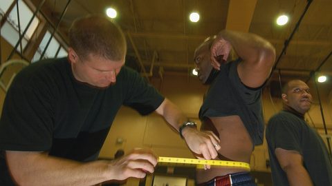
Waist circumference correlates with CT-measured visceral fat at r=0.6-0.8, depending on age, sex and ethnicity. That correlation is strong enough to make waist circumference a useful population screening tool. It is not strong enough to tell an individual how much of their abdominal girth is visceral versus subcutaneous fat. The same tape reading in two different people can represent very different ratios of visceral to subcutaneous fat.
Measurement protocol matters. WHO protocol directs you to measure at the midpoint between the lowest rib and the iliac crest. NIH/NHLBI protocol uses the top of the iliac crest. The difference can change the reading by 1-3 cm. Inter-observer error in untrained settings runs 0.5-2.5 cm. Self-measured waist circumference is unreliable, with errors commonly exceeding 3-5 cm, because people misunderstand the landmark and pull the tape too tight or too loose.
Waist Circumference Thresholds by Authority
The WHO (2000) defines increased metabolic risk at >94 cm for men and >80 cm for women, and substantially increased risk at >102 cm for men and >88 cm for women. These thresholds were derived from a Dutch population study by Lean et al. (1995). The NHLBI (ATP III, 2001) uses the same >102 cm/>88 cm thresholds for high risk. NICE CG189 (2022) adopts the WHO increased-risk thresholds of >94 cm/>80 cm. For Asian populations, the WHO Expert Consultation (2004) recommends thresholds of ≥90 cm for men and ≥80 cm for women, based on evidence that metabolic risk increases at lower waist circumferences in these populations.
Waist-to-Height Ratio Removes Sex-Specific Cut-Offs
NICE CG189 introduced waist-to-height ratio as a central adiposity marker alongside BMI, advising that the waist measurement should be less than half of height. The 0.5 threshold is a population heuristic, not a personalised risk score. The evidence comes from Ashwell et al. (2014), a systematic review of 31 studies covering approximately 300,000 adults. Waist-to-height ratio has the advantage of requiring no sex-specific cut-off, but it still cannot distinguish visceral from subcutaneous fat. A person with a waist-to-height ratio of 0.55 could have either high visceral fat or high subcutaneous fat or both. Only CT or MRI can tell you which.
What Waist Circumference Cannot Do: The Distinction Problem
The fundamental limitation of waist circumference and waist-to-height ratio is that they measure total abdominal girth, not the internal distribution of fat. Two people with a 100 cm waist can have very different amounts of visceral fat. One could store most of that girth in subcutaneous fat under the skin, with relatively little visceral fat around the organs. The other could have a smaller subcutaneous layer but substantial visceral fat pushing the waist outward from inside the abdominal cavity. The tape measure cannot tell the difference.
This is why some individuals with a normal waist circumference still have elevated cardiometabolic risk, and some with an elevated waist circumference have normal metabolic markers. The failure case is assuming that a waist circumference below a threshold guarantees low visceral fat. It does not. In clinical practice, waist circumference is a screening tool that flags people who need further assessment, just like BMI. It is not a diagnostic for visceral adipose tissue.
The Honest Caveat: No Single Number Tells You Your Visceral Fat Status
Waist circumference, waist-to-height ratio, and BIA estimates are all surrogates with individual error. CT and MRI are the only methods that directly measure visceral fat, and they are not available for routine screening. The thresholds that exist, the JASSO 100 cm², the WHO waist circumference bands, the NICE 0.5 rule, are all derived from specific populations and may not apply to you. The single thing that most often goes wrong is overconfidence: treating a waist circumference or a BIA reading as a precise measure of visceral fat when it is a rough estimate. Use these numbers as flags, not verdicts. If your waist-to-height ratio is above 0.5, ask a clinician whether you need further assessment. No tape measure can tell you what is happening inside your abdominal cavity.
Common Questions
Can I measure my own visceral fat at home?
No. Home BIA scales with a visceral fat estimate use proprietary algorithms that have an error of ±5-10% compared to CT or MRI. The reading changes with your hydration status, recent exercise and skin temperature. Do not treat it as a clinical measurement.
Is it possible to have high visceral fat with a normal BMI?
Yes. This is called normal-weight obesity. In NHANES data, a significant fraction of normal-BMI adults have excess body fat by DXA, and visceral fat can be elevated even when total body fat is normal. Waist circumference is a better screening tool than BMI for this scenario.
Can exercise specifically reduce visceral fat?
Visceral fat is more responsive to caloric deficit and exercise than subcutaneous fat. Moderate-to-vigorous aerobic exercise reduces visceral fat preferentially, but no exercise can target visceral fat alone. Overall fat loss is required.
What is the single best non-imaging measurement for visceral fat risk?
Waist-to-height ratio, with the NICE 0.5 threshold, is the best surrogate because it adjusts for stature and removes sex-specific cut-offs. But it is still a surrogate. Only CT or MRI can confirm visceral fat amount.